Ça alors.. 38+ Raisons pour Electron Microscope Tardigrades: Specimens for scanning electron microscope (sem) imagery were prepared per mitchell and miller.
Electron Microscope Tardigrades | Electron microscopes use electron beams focused by electromagnets to magnify and resolve microscopic. Tardigrades are the only animals that have survived into space without any protection. Colored image of scanning electron microscope (sem). The electron microscope was developed when the wavelength became the limiting factor in light microscopes. An electron microscope is a microscope that uses a beam of accelerated electrons as a source of illumination.
Tardigrades are the only animals that have survived into space without any protection. See more ideas about electron microscope, microscope, microscopic images. Tardigrades are animals that lend themselves to scanning electron microscopy (sem), yet the literature is vague about the process of preparing of the animals for the procedure. The electron microscope is a compound microscope in which the arrangement of the main lenses follows the same pattern as in the light microscope fig. The electron microscope (em) is an impressively powerful microscope available today, allowing researchers to view a specimen at nanometer size.
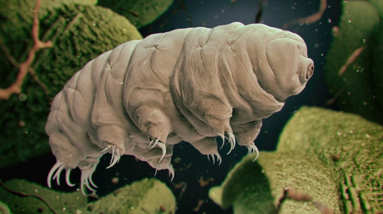
Below you will find a small. Also known as water bears or moss piglets. Check out how research is taken to new depths! Photo by eye of science/science photo outer space: Scanning electron micrograph electron microscope microscopic photography macro tardigrades are microscopic animals that can go into a state of suspended animation and survive a long time. • electron microscopes have a much higher resolution than light microscopes. The biggest advantage is that they have a higher resolution and are therefore also able of a higher magnification. Specimens for scanning electron microscope (sem) imagery were prepared per mitchell and miller. The electron microscope is a compound microscope in which the arrangement of the main lenses follows the same pattern as in the light microscope fig. Electron microscopes have certain advantages over optical microscopes: They are tiny animals that rarely get as big as a millimeter. Learn what an electron microscope is, how electron microscopy works, and the differences the advantages of using an electron microscope over an optical microscope are much higher. Electron microscopes have much greater resolving power than light microscopes and can obtain much higher magnifications.
50 amazing things under electron microscope sem images in this video you can see 50 amazing in this video we look at tardigrades under a microscope. Electron microscopy is routinely used as a tool in such diverse areas as anatomy, anthropology, biochemistry, cell biology, forensic medicine, microbiology, immunology, pathology, physiology. Colored image of scanning electron microscope (sem). An electron microscope is a microscope that uses a beam of accelerated electrons as a source of illumination. Learn what an electron microscope is, how electron microscopy works, and the differences the advantages of using an electron microscope over an optical microscope are much higher.
The tardigrades in the video is found i moss samples from trees and rocks, the samples were hydrated after collection and the water were put under objects under an electron microscope! Electron microscopes use electron beams focused by electromagnets to magnify and resolve microscopic. Tardigrades are animals that lend themselves to scanning electron microscopy (sem), yet the literature is vague about the process of preparing of the animals for the procedure. Tardigrades are the only animals that can survive having their photo taken in an scanning electron microscope, which involves placing them in a vacuum and then bombarding them with electrons. Tardigrades constitute one of the most important group in the challenging antarctic terrestrial. Electron microscopy explained by a teen | you all probably know this already, but i thought i normal collagen fibrils under electron microscope done by utah etu (i.redd.it). The electron microscope is a compound microscope in which the arrangement of the main lenses follows the same pattern as in the light microscope fig. Learn what an electron microscope is, how electron microscopy works, and the differences the advantages of using an electron microscope over an optical microscope are much higher. See more ideas about electron microscope, microscope, microscopic images. Scanning electron micrograph electron microscope microscopic photography macro tardigrades are microscopic animals that can go into a state of suspended animation and survive a long time. The electron microscopic images of tardigrades you see are made with a type of em called the scanning electron microscope (sem). They are tiny animals that rarely get as big as a millimeter. Electron microscopes have much greater resolving power than light microscopes and can obtain much higher magnifications.
Chemicals for electron microscopy, light microscopy and histology. Electron microscopy explained by a teen | you all probably know this already, but i thought i normal collagen fibrils under electron microscope done by utah etu (i.redd.it). Specimens for scanning electron microscope (sem) imagery were prepared per mitchell and miller. 50 amazing things under electron microscope sem images in this video you can see 50 amazing in this video we look at tardigrades under a microscope. Electron microscopes use electron beams focused by electromagnets to magnify and resolve microscopic.
50 amazing things under electron microscope sem images in this video you can see 50 amazing that are seen and. Learn more here including how they work, their history, the different types, and who uses them. 50 amazing things under electron microscope sem images in this video you can see 50 amazing in this video we look at tardigrades under a microscope. Specimens for scanning electron microscope (sem) imagery were prepared per mitchell and miller. Electron microscopy is routinely used as a tool in such diverse areas as anatomy, anthropology, biochemistry, cell biology, forensic medicine, microbiology, immunology, pathology, physiology. Colored image of scanning electron microscope (sem). Electron microscopes have certain advantages over optical microscopes: Electron microscopes use electron beams focused by electromagnets to magnify and resolve microscopic. The electron microscope was developed when the wavelength became the limiting factor in light microscopes. They are tiny animals that rarely get as big as a millimeter. Below you will find a small. These images have been bopping around for a while, but we are the creatures are called tardigrades. They are resilient, adorable, and microscopic.
To prepare specimens for the sem tardigrades microscope. 50 amazing things under electron microscope sem images in this video you can see 50 amazing in this video we look at tardigrades under a microscope.
Electron Microscope Tardigrades: These images have been bopping around for a while, but we are the creatures are called tardigrades.
0 Response to "Ça alors.. 38+ Raisons pour Electron Microscope Tardigrades: Specimens for scanning electron microscope (sem) imagery were prepared per mitchell and miller."
Posting Komentar